Cranial Module
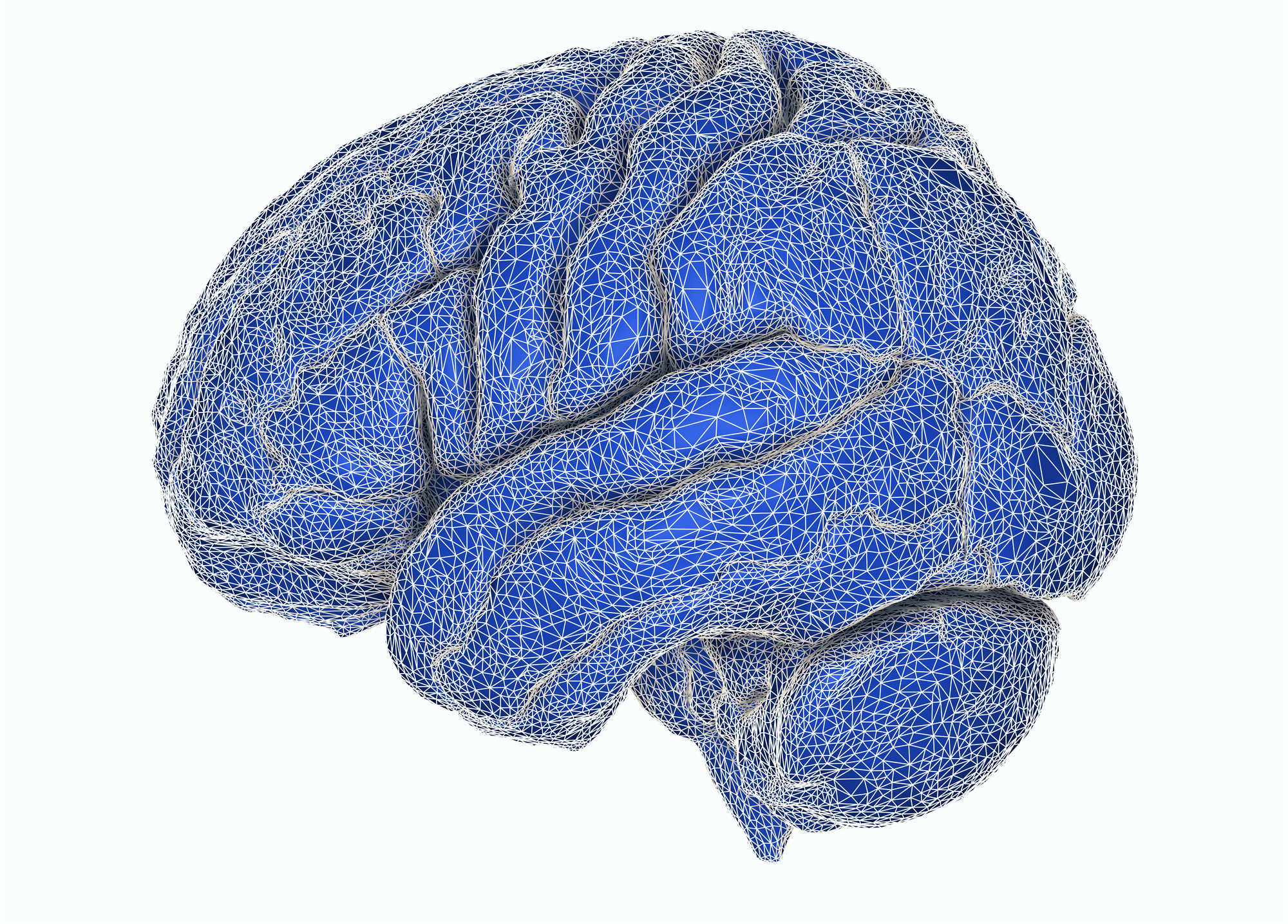
Build a surgical case from multimodal neuroimaging
Bring CT, MRI, vascular imaging, landmarks and lesions into one consistent patient model.
ImFusion Cranial provides the SDK building blocks to segment, register, organize, and serve this information across planning and navigation workflows.
One coherent patient model, ready for navigation workflows
Registration results, segmentations, meshes, and landmarks remain associated with the same named patient anatomy. This allows planning views, image fusion, measurements, visualization, and navigation workflows to operate on a shared representation of the case.
Build the anatomy model
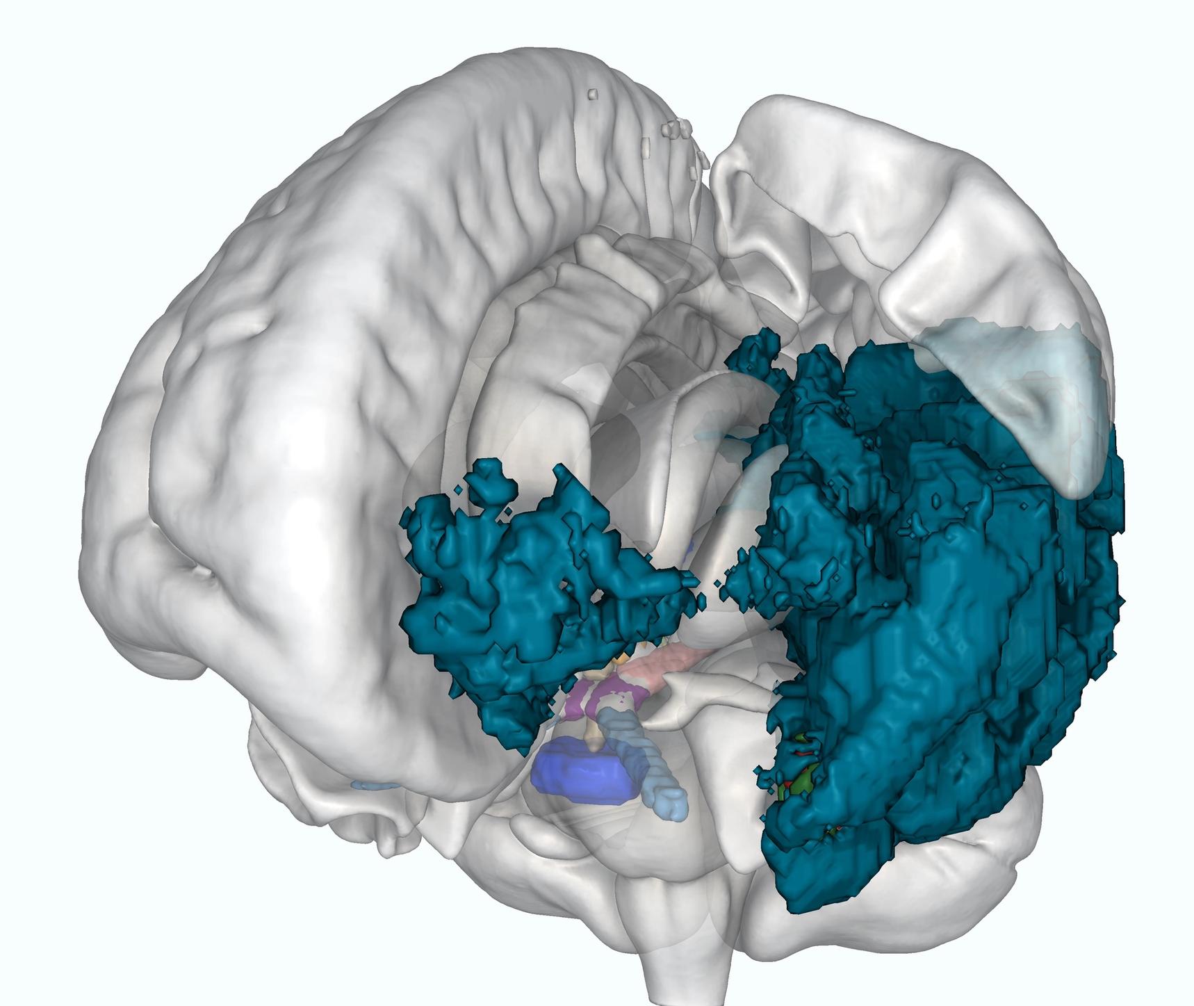
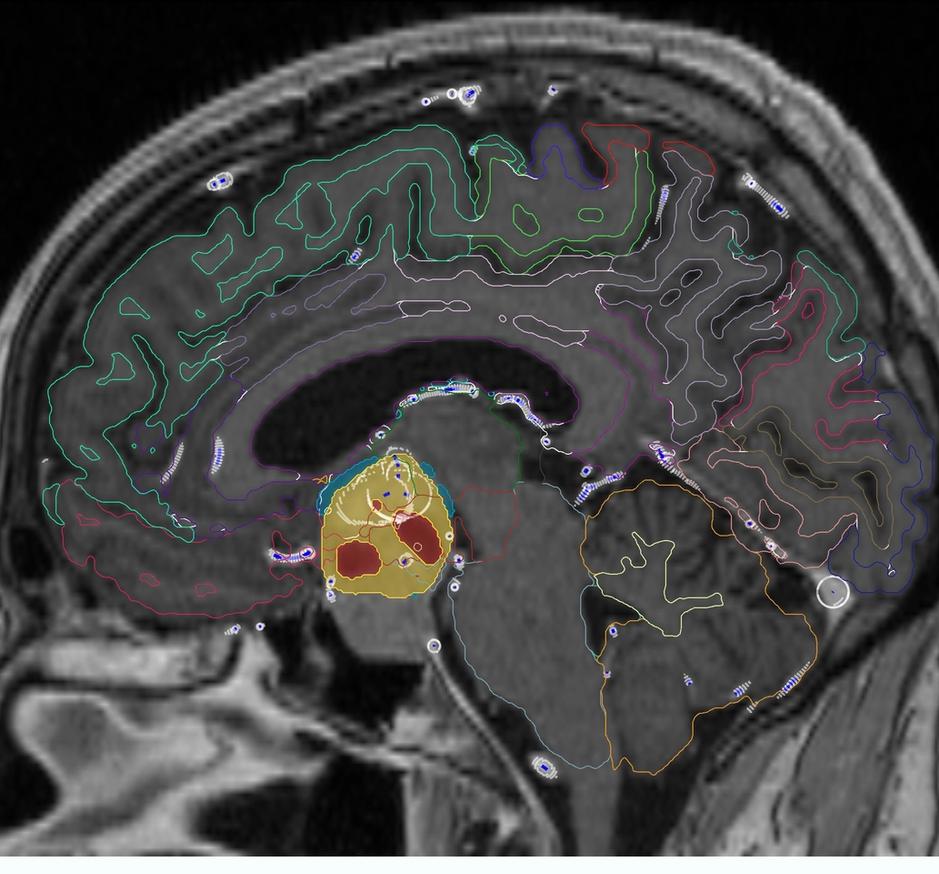
ImFusion Cranial converts patient imaging into a structured anatomical model for planning, registration, and visualization. It combines preprocessing and segmentation of cranial anatomy, brain structures, and tumors, while keeping derived structures associated with the source imaging.
- Segment skull and craniofacial bone from CT
- MRI bias field correction and skull stripping
- Cortical and subcortical brain structures from T1-weighted MRI
- Multi-contrast tumor segmentation on any or all of T1, T1CE, T2, and FLAIR
Map vascular anatomy
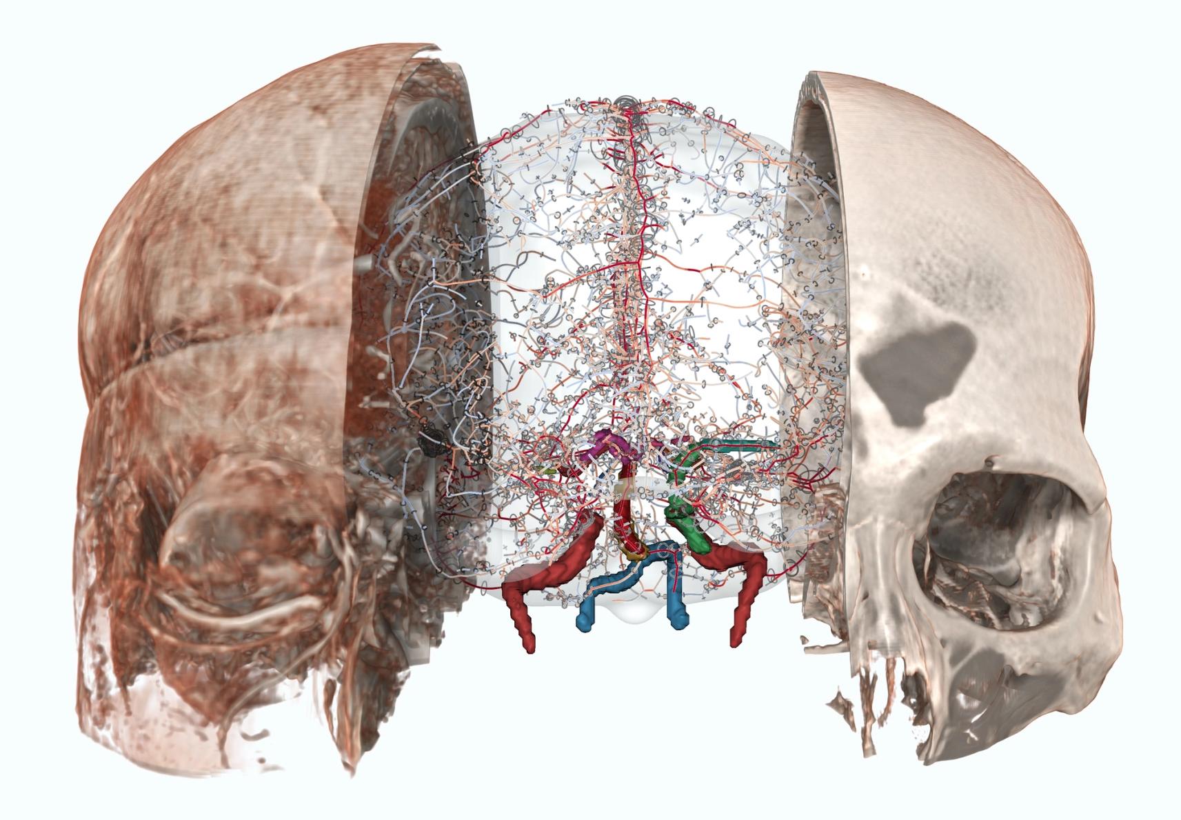
Vascular structures can be incorporated into trajectory planning, corridor selection, tumor assessment, and multimodal visualization. Cranial supports general cerebral vessel segmentation on T1CE MRI, ToF-MRA, and CTA, plus dedicated arterial models for the Circle of Willis and extra-cranial brain arteries.
- Vessel trees represented as graphs
- Centerline and vessel-diameter measurements
- Visualization of vessels together with the surrounding anatomy
Align modalities and reference frames
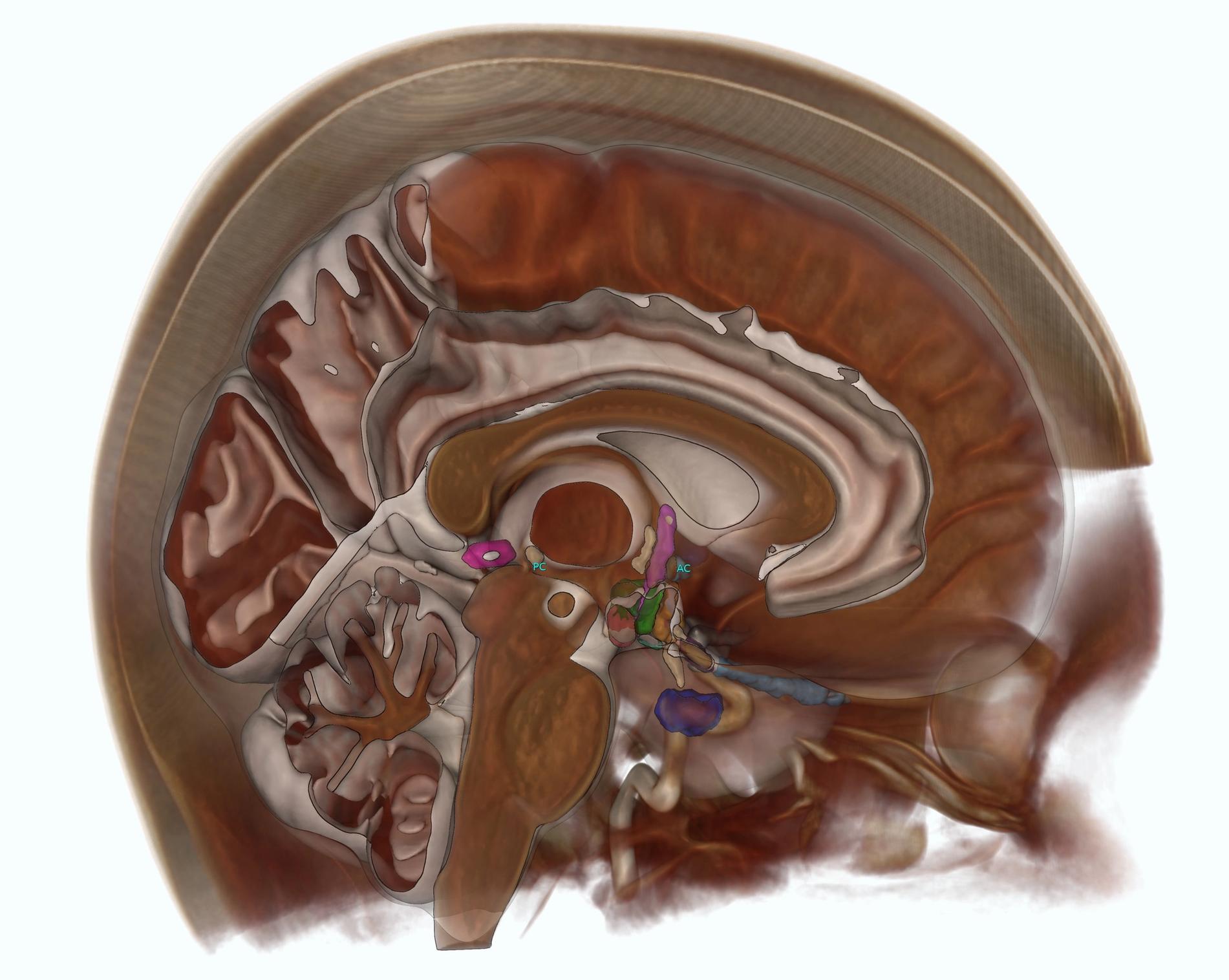
ImFusion Cranial places patient anatomy, vascular structures, and image data in a shared spatial reference. AC-PC detection defines an anatomical coordinate system on T1-weighted, T1CE, T2-weighted, or FLAIR MRI, while multi-volume registration aligns CT, MRI, vascular imaging, and longitudinal follow-up studies. Segmentations, meshes, and landmarks remain associated with the registered images.
- AC-PC landmarks and anatomical coordinate system
- CT-MRI, MRI-MRI, and longitudinal registration
- Propagation of segmentations, meshes, and landmarks with registered images
Build application-specific planning workflows
Clinical applications rarely follow a single imaging pipeline. For DBS planning, stereotactic targeting, craniotomy planning, and navigation, Cranial provides patient-specific anatomy, registration, landmarks, vessels, and real-time visualization as configurable building blocks. The resulting workflow can reflect your imaging inputs, planning steps, display conventions, and handoff to guidance.
- Define targets, risk structures, and access corridors in one patient model
- Anchor planning, display atlas structures, or patient-specific anatomy
- Combine CT bone, MRI soft tissue, vessels, tumors, landmarks, and planned objects
- Integrate the resulting case into your planning or navigation application
Get a technical walkthrough of the platform to see our key workflows in action and assess fit.
Product Demo
In a product demo, we show you how the ImFusion Suite, SDK, and ImFusion Labels support medical imaging and data annotation workflows. We focus on the capabilities relevant to your application and discuss directly how our technology could fit into your product or support your use case.
- See integration points mapped to your specific tech stack
- Identify bottlenecks before committing development resources
- Validate data format compatibility and transformation requirements
- Get immediate answers from engineers who built the system